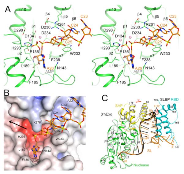
Figure 3.
The 3′ flanking sequence of the SL RNA is located in the 3′hExo active site. (A). Interactions between the 3′ flanking sequence of the SL RNA (orange) with the active site of 3′hExo nuclease domain (green). The bound positions of AMP (gray) and two metal ions (pink spheres) to the nuclease domain of 3′hExo as observed earlier are also shown (20). (B). Molecular surface of the active site region of 3′hExo colored based on electrostatic potential. The SL RNA is shown as a stick model (orange). The black arrow indicates another opening from the active site, through which 3′hExo may accommodate longer RNA molecules. (C). Overlay of the structures of the ternary (SL-SLBP-3′hExo, in color) and binary (gray for 3′hExo and black for SL) complexes. The superposition is based on the nuclease domain of 3′hExo.

