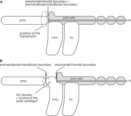
Fig. 4.

Diagram to explain the question regarding the nature of the polar cartilage. (A) It is generally accepted that the rostral, prechordal part of the neurocranium is formed of premandibular ectomesenchyme. Thus, the boundary between the premandibular and mandibular ectomesenchyme is expected to be found at the same level as the boundary between the prechordal and chordal cranium, and the polar cartilage is simply understood as the posterior part of the trabecula. In this scheme, the hypophysis is found between the caudal portions of trabeculae, the prechordal cranial element. (B) If the polar cartilage represents the ‘prechordal’ mandibular arch-derivative, we will have to assume the presence of an enigmatic ectomesenchymal domain corresponding to the ‘HC domain’. This diagram is based on the understanding of Haller (1923) and Allis (1938) shown in Fig. 5A. In this scheme, the hypophysis represents the boundary between the ectodermal portions covering the forebrain and the pharynx. hc, hyoid arch crest cells; mnc, mandibular arch crest cells; nt, notochord; pch+orb, parachordal and orbital cartilages; pmc, premandibular crest cells.
