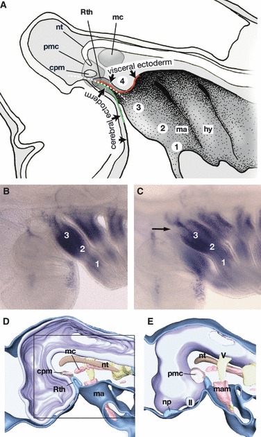
Fig. 5.

Topographical relationship between the mandibular arch and the hypophyseal anlage. (A) A medial view of the early pharyngula of a shark. Redrawn from Haller (1923). According to Haller, Rathke’s pouch does not arise as a part of the oral ectoderm, which is not defined at the stage of initial hypophyseal development, but at the junction of two different ectoderms: the cerebral ectoderm that covers the forebrain, and the visceral ectoderm that covers the mandibular arch and forms the stomodaeum. Haller (1923) divided the mandibular arch along the dorso-ventral axis into four subdivisions (1–4), termed, from a ventral to a dorsal direction, ‘Unterkieferstück (lower jaw region)’ (1), ‘Zwischenstück (intermediate or jaw joint region)’ (2), ‘Oberkieferstück (maxillary region)’ (3), and ‘Kieferaugenspaltstück (hypophyseal cushion region, abbreviated as HC region in the text)’ (4). The HC region is found just caudal to the hypophyseal anlage, and the polar cartilage is suggested to develop in this domain. Thus, Allis (1938) regarded the polar cartilage as the mandibular arch derivative. (B) Dlx1 expression pattern in a pharyngula of Scyliorhinus torazame. Lateral view. (C) The same embryo as B, slightly tilted to view the embryo ventrally. The HC region of this embryo does not appear to express Dlx1. (D,E) Partially reconstructed early pharyngula of an elephant fish, Callorhynchus milii. Medial (D) and lateral (E) views (E is left–right inverted for comparison). Square in (D) indicates the equivalent region shown in (A). These figures show that the embryonic pattern is highly conserved in chondrichthyans and lamprey, in which nasal and hypophyseal placodes are more closely associated with each other. cpm, Commissure part of the premandibular cavity; hy, hyoid arch; II, optic nerve; ma, mandibular arch; mam, mandibular arch mesoderm; mc, mandibular cavity; np, nasal placode; nt, notochord; pmc, premandibular cavity; Rth, Rathke’s pouch; V, trigeminal nerve anlage.
