
Fig. 5.
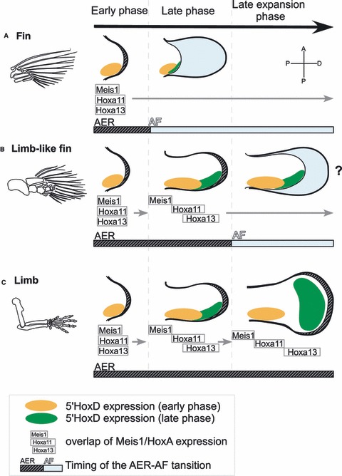
The repression mode of the AF. Morphological differences among fins, limb-like fins, and limbs are explained by a combination of developmental mechanisms: separation of the Meis/HoxA11/HoxA13 expression domains, the degree of 5′HoxD late expansion, and the occurrence and timing of the AER–AF transition. (A) In fin development in actinopterygians, the AER–AF transition occurs at early stages of development, and fin mesenchyme starts differentiating into endoskeleton before the completion of successive change in the gene expression domains (Grandel & Schulte-Merker, 1998). With this timing of transition, the Meis/Hoxa11/Hoxa13 expressions overlap within a domain, and the late phase of 5′Hoxd regulation is not functional (Sordino et al. 1995; Grandel et al. 2000; Metscher et al. 2005). (B) In the limb-like fin development of sarcopterygian fish, the AER–AF transition is speculated to occur later than in other fish (Thorogood, 1991). As a result, distinct Meis/HoxA11 expression domains occur, resulting in formation of the stylopod. When the AER–AF transition occurs, the separation of the HoxA11/HoxA13 domains is still incomplete, and the 5′HoxD domain is posteriorly restricted (Shubin et al. 2009; Woltering & Duboule, 2010; Schneider et al. 2011), resulting in the formation of a zeugopod and dwarfish autopod without any digits (Cote et al. 2002; Shubin et al. 2006; Boisvert et al. 2008). (C) In the limb development of tetrapods, the AF does not form, and the sustained AER promotes the proliferation of undifferentiated mesenchyme (Guo et al. 2003). The expression domain of HoxA13 is separated from that of HoxA11 (Yokouchi et al. 1991; Nelson et al. 1996; Stadler et al. 2001; Sato et al. 2007) and the late-phase 5′HoxD domain is expanded along the AP axis, giving rise to a complete set of autopod elements, including digits. In (A) and (B), the AF formed after the AER–AF transition represses any further progression of molecular mechanisms in the endoskeletal region and discontinues the PD and AP patterning therein. Distal is to the right; anterior is to the top.