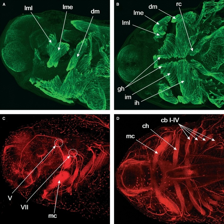
Fig. 1.

Whole-mount immunolabeled stage 41 Ambystoma mexicanum embryo. Max-projections of CLSM-image stacks. (A) Lateral view of the head. Desmin is used as a muscle marker and is expressed in the differentiating cranial muscles. The levator mandibulae longus (lml) is extending its origin dorsally and the levator mandibulae externus (lme) is extending caudally. The depressor mandibulae muscle (dm) is extending close to its insertion. (B) Ventral view of the head. Desmin expression in the differentiating cranial muscles. In addition to the muscles visible in lateral view, we now also see ventral muscles such as the intermandibularis (im) and interhyoideus (ih) muscles, and how they insert on the ventral midline. Also visible are the hypobranchial muscles geniohyoideus (gh) and rectus cervicis (rc). (C) Lateral view of the head. Nerve axons are marked by antibodies towards acetylated alpha-tubulin, cartilage is marked by a collagen II antibody. A comparison with (A), which shows the same specimen viewed in another channel to show the muscle staining, shows that the levator muscles are innervated by cranial nerve V and insert on the dorsal side of Meckel’s cartilage (mc), whereas the depressor mandibulae is innervated by cranial nerve VII and inserts at the caudal end of Meckel’s cartilage. (D) Markers as in (C). Ventral view of the head. By comparing with (B), we can see how cartilages and muscles match up. Levator and depressor mandibulae muscles, for example, attach to Meckel’s cartilage (mc), and the interhyoideus muscle (ih) to the ceratohyal (ch). Gill muscles are associated with the ceratobrachials I–IV (cb I–IV).
