
Fig. 1.
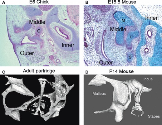
Middle ear ossicles in mammals and birds. (A) Frontal section through the developing middle ear of a chick showing the columella (c) spanning the gap between the external and internal ear at E (embryonic day) 6. (B) Sagittal section through the developing murine middle ear showing three ossicles, the malleus (M), incus (I) and stapes (S), between the external and inner ear at E15.5. (C,D) MicroCT images. (C) Footplate of the columella (c) inserting into the oval window of the inner ear in an adult partridge. The shaft and footplate of the columella are ossified while the extracolumella arms, which interact with the tympanic membrane, remain cartilaginous and are not picked up by microCT. (D) Three ossicles form a chain in a P (postnatal day) 14 mouse.