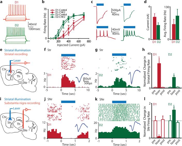
Figure 2. ChR2-mediated excitation of direct- and indirect-pathway MSNs in vivo drives activity in basal ganglia circuitry.
(a) Whole-cell current-clamp recordings from ChR2-YFP+ neurons in vitro demonstrate normal current-firing relationships consistent with D1-MSNs (red traces) or D2-MSNs (green traces) (D1-Control, n=10; D1-ChR2, n=3; D2-Control, n=7; D2-ChR2, n=3). (b) Firing rate plotted as a function of injected current in D1-MSNs or D2-MSNs expressing either GFP or ChR2-YFP. (c) ChR2-mediated photocurrents (top) and spiking (bottom) in D1 (left) and D2 (right) MSNs. In this and subsequent panels, blue bars indicate illumination time. (d) Summary of ChR2-mediated photocurrents (left) and spiking (right) for D1-ChR2 (n=5) and D2-ChR2 (n=4) cells. (e) Schematic of in vivo optical stimulation and recording in the striatum (Str). Cortex (Ctx), thalamus (Th), substantia nigra pars reticulata (SNr). (f) An example MSN recorded from the striatum of an anesthetized D1-ChR2 mouse that displayed increased firing in response to illumination. Insets in f-g and j-k show spike waveform with illumination (blue) or without illumination (grey). Scale bar applies to insets in f-g and j-k. (g) An example of a light-sensitive MSN from a D2-ChR2 mouse. (h) Normalized change in MSN firing rates in response to striatal illumination in D1-ChR2 (n=16) or D2-ChR2 (n=10) mice. (i) Schematic of in vivo optical stimulation in striatum and recording in SNr. (j) An example of a SNr neuron recorded from a D1-ChR2 mouse that was inhibited by direct pathway activation. (k) An example of a SNr neuron recorded from a D2-ChR2 mouse that was excited by indirect pathway activation. (l) Normalized change in SNr firing rate in response to activation of the direct (D1, n=8) or indirect (D2, n=4) pathways. Error bars are SEM.

