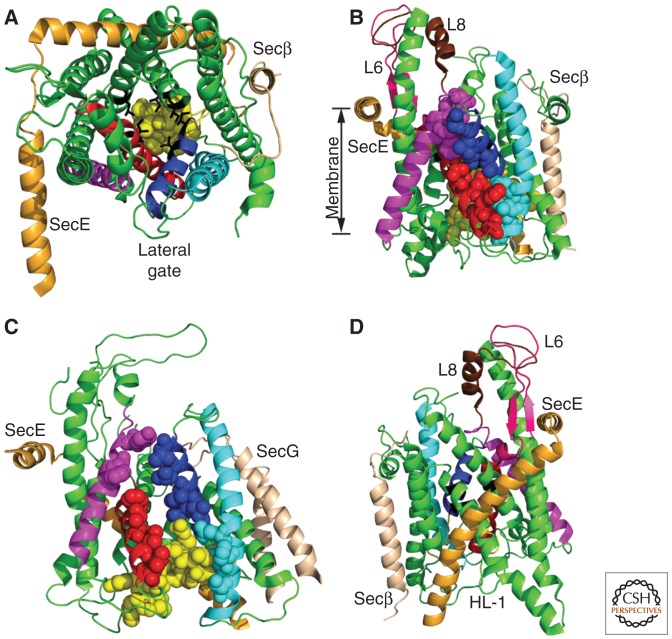
Figure 2.
SecYEβ and SecYEG translocation channels. TM spans of SecY are color coded as follows: TMs 1, 4–6, 9–10 (green), TM2 (blue), TM3 (cyan), TM7 (red), and TM8 (magenta). (Yellow spheres) The plug domain. Cytosolic loops 6 and 8 are pink and chocolate, respectively, in panels B and D. (A) The cytosolic face of the Methanocaldococcus jannaschii SecYEβ complex in the closed conformation. (Black sticks) Pore ring residues. (B) Lateral gate of M. jannaschii SecYEβ viewed from the plane of the membrane. (Spheres) Lateral gate contact residues (LGCRs). (C) The partially open conformation of the Thermotoga maritima SecYEG complex. (D) The hinge domain of M. jannaschii SecYEβ. The HL-1 hinge loop is labeled. All structure views were generated using PyMOL and PDB files 1RHZ and 3DIN.

