Figure 3.
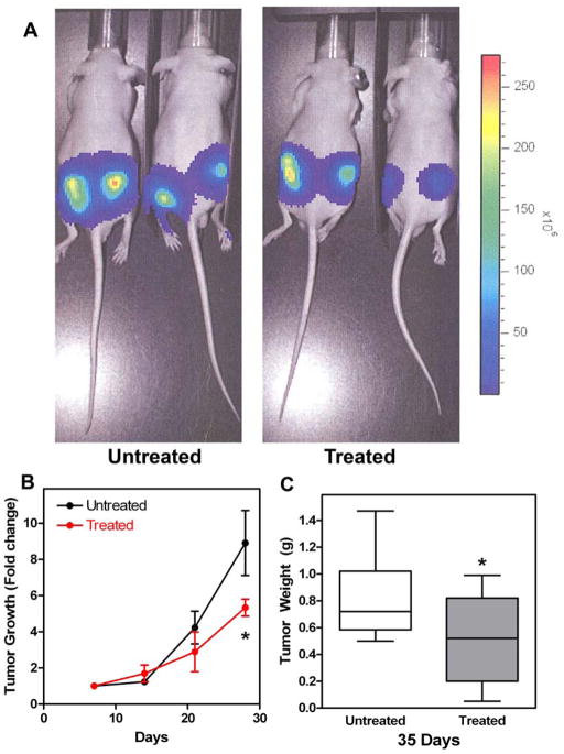
Panel A. Representative bioluminescence imaging of tumors in untreated and treated nude mice. Animals were imaged on days 7, 14, 21 and 28. The figure is from day 28. Panel B. FM19 delayed tumor growth in treated nude mice. Mice were prepared for non-invasive bioluminescence imaging during treatment with FM19 in the drinking water (3 mg/ml). The data were normalized to day 7 for each injection site and the fold change for each subsequent scan is plotted. Error bars indicate standard deviation from 4 (untreated) or 5 (treated) animals each with 2 injections. Panel C. FM19 reduced the overall tumor weight in nude mice. Mice were treated for with FM19 in the drinking water (3 mg/ml) for 5 weeks, tumors were excised and weighed. Error bars represent the standard deviation of 8 (untreated) or 10 (treated) tumors.

