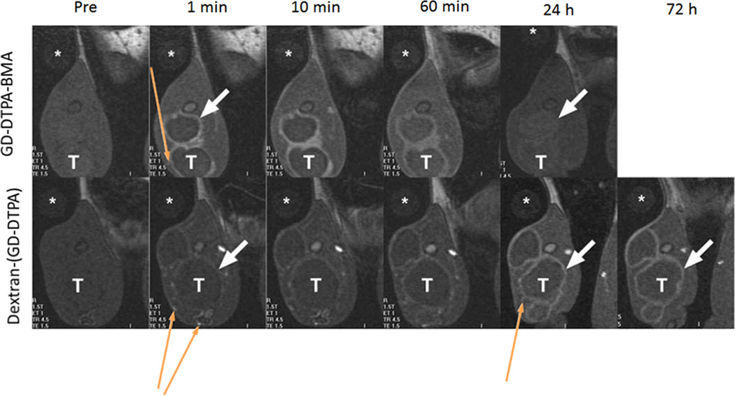
Figure 4.
Coronal T1-weighted gradient echo images of VX2 tumor bearing rabbits at 1.5T showing tumor rim (white, short, thick arrow) and peritumoral vessels (yellow, thin, long arrows) before and at 1, 10 and 60 minutes and 24 and 72 hours after iv injection of Gd(DTPA-BMA) (Omniscan®) and Dextran-(Gd-DTPA) at 0.1 mmol-Gd kg−1. (*, water phantom; T, tumor). Adapted from ref. 39.

