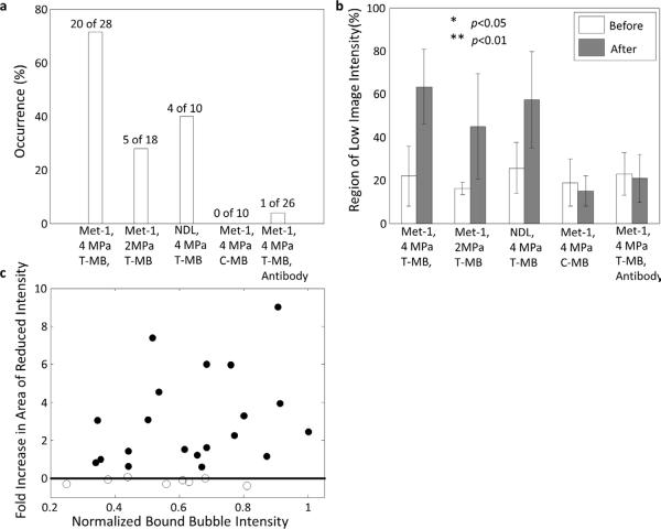
Figure 4.
The occurrence and area of regions with reduced contrast image intensity in Met-1 and NDL tumors with two acoustic pressures with data combined between cRGD and LXY3-conjugated microbubbles. (a) Using 4 MPa-PNP pulses, regions of reduced flow were observed in 71% of 28 Met-1 and 40% of 10 NDL tumors. Using 2 MPa-PNP pulses, regions of reduced flow were observed in 28% of 18 Met-1 tumors. Using 4 MPa-PNP pulses to destroy flowing non-targeted microbubbles, regions of reduced flow were not observed in 10 Met-1 tumors. With pre-treatment by anti-CD41 antibody, reduced blood flow was produced in 1 of 26 insonified Met-1 tumors (4%). (b) The area with reduced contrast intensity before and after bound microbubble destruction. For a 4 MPa pulse pressure, the area of reduced flow increased from 22 ± 13% to 63 ± 17% (p<0.01) in Met-1 tumors and 16 ± 3% to 45 ± 24% (p<0.01) in NDL tumors by destruction of bound targeted microbubbles. For Met-1 tumors and a 2 MPa pulse pressure the area with reduced flow increased from 25 ± 13% to 57 ± 22% (p<0.05). With the destruction of flowing non-targeted microbubbles, the area of reduced contrast intensity was not significantly increased. Similarly, in the inhibition study, the area of regions with reduced contrast intensity was unchanged when platelet adhesion was blocked. (c) The increased area with low contrast image intensity before and after microbubble destruction was independent of the echo intensity generated by bound microbubbles within the local region.

