
Figure 6.
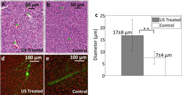
Histological and confocal results for the insonation of bound microbubbles. (a, b) H&E of insonified (a) and control (b) Met-1 tumors from mice that had been injected with LXY-3-conjugated microbubbles. (c) Summary of vessel diameter assessed from H&E, combining studies with Met-1 and NDL tumors and LXY-3 and RGD-conjugated microbubbles. The mean diameter of vessels was greater in the ultrasound-treated tumors (17 ± 8 μm) as compared with control tumors (7 ± 4 μm). (d, e) Confocal images from control and insonified tumors from mice that had been injected with LXY-3-conjugated microbubbles. FITC-Dextran (green) overlaid on bright field image (red). Insonified tumors (d) include unevenly distributed FITC-dextran which has extravasated. In control tumors (e), discrete blood vessels are visualized. ** indicates p<0.01 (e). Scale bar in a and b represents 50 μm; while scale bar in d and e represents 100 μm.