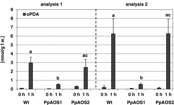
Figure 8.

Analysis of cis(+)-OPDA in P. patens WT, ΔPpAOS1 and ΔPpAOS2.cis(+)-OPDA was extracted from unwounded (control) and wounded (1h after wounding stimulus) moss by employing the methyl-tert-butyl ether technique [41] and analyzed via LC/MS [42]. Shown are the results from two independent experimental datasets. Data are presented as mean values with standard deviations from two - six biological replicates. Values with significant differences (Students T-Test; P < 0.05) are indicated above the respective column by different letters.
