
Figure 3.
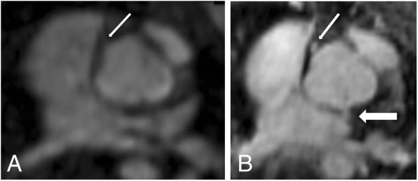
11-year-old patient after surgical correction of a common arterial trunk (same patient as in Figure2). Axial plane through the ascending aorta at the level of the coronary ostia in FP-MRA (A) and HR-MRA (B). Whereas in the FP-MRA only the origin of the right coronary artery can be detected (thin arrow), the HR-MRA accurately depicts the origins of both coronary arteries (thin arrow: right coronary artery; thick arrow: left coronary artery) revealing an abnormal origin of the left coronary artery out of the non-coronary cusp.