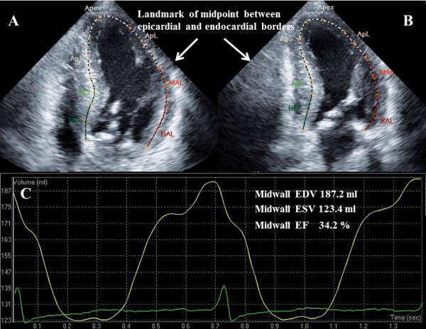
Figure 1.

Examples of midwall EF measurements. (A) 4-chamber view. (B) 2-chamber view. The positioning of the midwall is determined by the landmark of the midpoint between the epicardial and endocardial borders. (C) Examples of midwall volume curve using speckle tracking method. EF = ejection fraction. EDV = end diastolic volume; ESV = end systolic volume.
