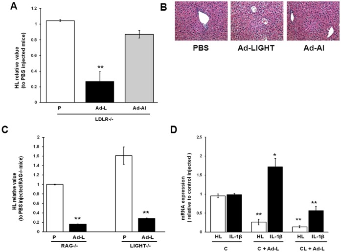
Figure 2. Decreased HL expression in Ad-LIGHT infected mice is independent of T cells and IL-1β.
Mice were injected with PBS or adenoviral vectors (1.25×10⁹ pfu/mouse) and sacrificed on the 7th day. (A) Liver HL mRNA expression in LIGHT adenovirus (Ad-L), human apoA-I adenovirus (Ad-AI) or PBS (P) injected LDLR−/− mice was analyzed by real time PCR. (B) Morphology of liver from PBS and adenoviral infected mice (H & E staining, 20x objective). (C) Liver HL mRNA expression in Ad-L and P injected RAG−/−LDLR−/− and LIGHT−/−LDLR−/− mice. (D) Liver HL and IL-1β mRNA expression in Ad-L injected LDLR−/− mice treated with control (C) or clodronate (CL) liposomes. The virus was injected 2 days after clodronate injection. (n = 3; *p<0.05, **p<0.01; for panels A and C: vs. PBS treated mice; for Panel D: vs. control liposome treated mice.).

