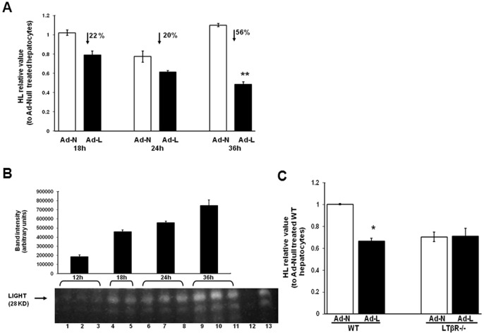
Figure 4. LIGHT and HL expression in Ad-LIGHT transduced primary hepatocytes in vitro.
(A) HL mRNA expression in Ad-LIGHT (Ad-L) and Null adenovirus (Ad-N) infected primary hepatocytes from wild type mice 18, 24 and 36 hours post-infection. The numbers indicate the % decrease in expression in the Ad-LIGHT infected cells relative to the Ad-N infected cells. (B) Total protein from 1×10⁶ Ad-LIGHT infected hepatocytes (in triplicate) at various times post-infection were immunoblotted with anti-mouse LIGHT antibody. Lane 12 is from non-infected hepatocytes and lane 13 is from Tg-LIGHT mouse spleen. (C) Liver HL mRNA expression in Ad-N and Ad-L infected hepatocytes from LDLR−/− and LTβR−/−LDLR−/− mice. (n = 3; *p<0.05, **p<0.01 vs. Ad-N).

