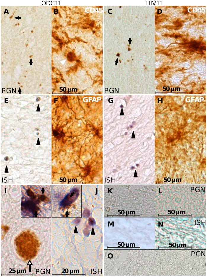
Figure 3. Bacterial detection in human brain.
Autopsy-derived ODC (A), and HIV (C) brain specimens were immunolabeled with anti-peptidoglycan antibody. Peptidoglycan (PGN)-positive bodies (arrows) were morphologically consistent with bacteria and smaller than CD45 immunopositive microglia from ODC (B) and HIV (D) patients, imaged at the same magnification. Double DIG-labeled EUB 338 probe in situ hybridization (ISH) against the 16 s rRNA gene was hybridized with slides from the same ODC (E) and HIV (G) patients and labeled with alkaline phosphatase-conjugated sheep anti-DIG FAB` fragments and stained with NBT/BCIP. ISH-positive bodies featured morphology resembling bacteria (arrow heads) and were smaller than GFAP immunopositive astrocytes in ODC (F) and HIV (H) patient sections. Peptidoglycan-labeled cells with both spherical and rod morphology were observed within the brain parenchyma and in a blood vessel (I) (White arrow) of an ODC patient. Peptidoglycan immunopositive bodies were observed within the cytoplasm of GFAP-immunolabeled astrocytes (I, inset) and Iba-1 immunolabeled microglia (J, inset). Spherical clusters of EUB 338 hybridized cells J) were evident (black arrowheads). Slides from the same ODC11 (K) and HIV11 (L) patients were processed under identical conditions except that the primary antibody was omitted. A scrambled DIG-labeled probe was hybridized to slides from the ODC11 (M) and HIV11 (N) under identical conditions used for the EUB 338 probe. In all cases specific signals were not detected. (Original magnification 200×). (O) A section from the forebrain of one of the FIV-infected cats was immunostained with the anti-PGN antibody and developed with DAB with no detectable signal. (Bar: A–H, 50 microns; I, 25 microns; J, 20 microns; K–N, 50 microns) (Magnification: A–H, 200×; I, 400×; J, 600×; I and J insets, 600×).

