
Figure 1.
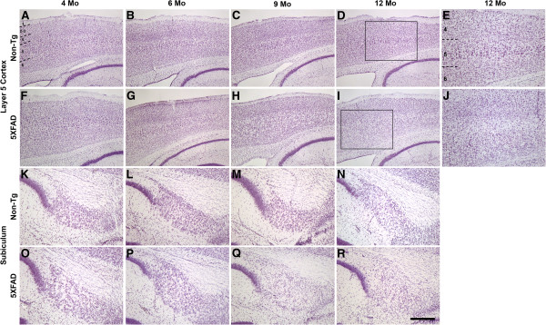
5XFAD mice exhibit neuron loss in Layer 5 cortex and subiculum. Parasagittal brain sections from representative 4- to 12-month-old non-transgenic littermate (A-E, K-N) and 5XFAD (F-J, O-R) female mice were stained with cresyl violet and micrographed to image Layer 5 cortex (A-J) and subiculum (K-R). Numbers indicate cortical layers and dashed lines identify borders between layers. Boxes in D and I outline the areas of increased magnification shown in E and J, respectively. The 5XFAD mouse exhibits visible loss of large pyramidal neurons at 9 months of age as evident by a decrease in stained neurons (H, Q, I, R, J). Increased magnification (J) shows a clear loss of large pyramidal neurons in Layer 5 of the 5XFAD mouse at 12 months compared to the non-transgenic (E). Scale bar in R = 280 μm for A-D, F-I; 110 μm for E, J; 80 μm for K-R.