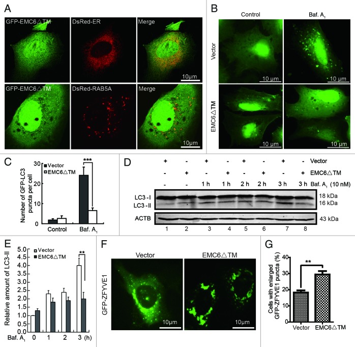
Figure 8. EMC6ΔTM suppresses cell autophagy. (A) U2OS cells were cotransfected with plasmids expressing GFP-EMC6ΔTM and DsRed-ER or DsRed-RAB5A for 24 h and observed by confocal microscopy. (B) Representative fluorescence microscopy images obtained from U2OS cells cotransfected with GFP-LC3 and vector or EMC6ΔTM for 30 h and treated with or without 10 nM bafilomycin A1 (Baf. A1) for the last 4 h. (C) Quantification of GFP-LC3 dots in control or EMC6ΔTM-expressing cells treated with reagents as indicated in (B). Results are means ± SD of at least 100 cells scored (***p < 0.001). (D) U2OS cells were transfected with vector or EMC6ΔTM for 24 h, and incubated with or without 10 nM Baf. A1 for the indicated times. The levels of endogenous LC3-I and LC3-II were analyzed by western blot. (E) Quantification of the amounts of LC3-II relative to ACTB treated as in (D). The average value in the vector-transfected cells without Baf. A1 treatment was normalized as 1. Data are the means ± SD of results from three experiments (**p < 0.01). (F) U2OS cells were cotransfected with GFP-ZFYVE1 and EMC6ΔTM or vector for 24 h and observed by fluorescence microscopy. (G) Numbers of cells with enlarged GFP-ZFYVE1 structures were quantified (means ± SD) by scoring at least five random fields. **p < 0.01.

An official website of the United States government
Here's how you know
Official websites use .gov
A
.gov website belongs to an official
government organization in the United States.
Secure .gov websites use HTTPS
A lock (
) or https:// means you've safely
connected to the .gov website. Share sensitive
information only on official, secure websites.
