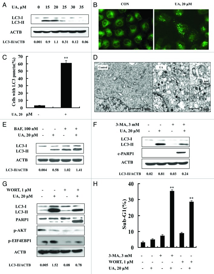
Figure 2. UA induced cytoprotective autophagy in MCF-7 human breast cancer cells. (A) UA caused an increased LC3-I to LC3-II conversion analyzed by western blotting (24 h). (B) Puncta distribution of LC3 induced by UA detected by immunofluorescence staining (24 h). (C) Quantitative analysis of LC3 puncta in cells in UA-treated and untreated cells. (D) TEM analysis of autophagic vacuoles in UA-treated and untreated cells (12 h). E. Increase of autophagosome formation was involved in UA-induced autophagy in MCF-7 cells. The cells were treated with 20 μM UA for 24 h in the presence or absence of 100 nM BAF (added 2 h before cell harvest) and then LC3 was analyzed by western blotting. (F and G) Effects of autophagy inhibitor 3-MA (F) or WORT (G) on UA-induced LC3-I to LC3-II conversion and PARP1 cleavage examined by western blotting. (H) Effects of autophagy inhibitor 3-MA or WORT on UA-induced apoptosis. The cells were treated with 20 μM UA in the presence or absence of 3-MA (3 mM) or WORT (1 μM) for 24 h and then apoptosis was assessed, measured by sub-G1 analysis (n = 3, **p < 0.01). (The blots shown are representative of three independent experiments).

An official website of the United States government
Here's how you know
Official websites use .gov
A
.gov website belongs to an official
government organization in the United States.
Secure .gov websites use HTTPS
A lock (
) or https:// means you've safely
connected to the .gov website. Share sensitive
information only on official, secure websites.
