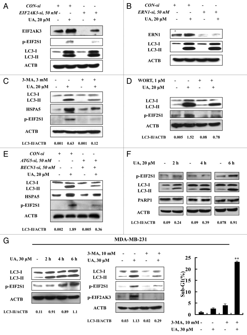
Figure 4. ER stress was an effect rather than a cause of UA-induced autophagy. (A and B) Effects of EIF2AK3 (A) or ERN1 (B) knockdown on UA-induced LC3-I to LC3-II conversion. The cells were transfected with 50 nmol/L of EIF2AK3 siRNA or 50 nmol/L of ERN1 siRNA using siPORT™ NeoFX™ Transfection Agent. At 24 h post-transfection, the cells were treated with 20 μM for 24 h and then EIF2AK3, p-EIF2S1, ERN1 and LC3 were assessed by western blotting. (C and D) Effects of autophagy inhibition by 3-MA (C) or WORT (D) on UA-induced HSPA5 expression and EIF2S1 phosphorylation. The cells were treated with 20 μM UA in the presence or absence of 3-MA or WORT for 24 h and were analyzed by western blotting. (E) Effects of autophagy inhibition by BECN1/ATG5 knockdown on UA-induced HSPA5 expression and EIF2S1 phosphorylation. The cells were simultaneously transfected BECN1 and ATG5 siRNAs using siPORT™ NeoFX™ Transfection Agent. After 24 h transfection, the cells were treated with 20 μM UA for 24 h and were assessed by western blotting. (F) Time-course analysis of key parameters of ER stress, autophagy and apoptosis induced by UA in MCF-7cells. The cells were treated with UA for the indicated time and then LC3, EIF2S1 phosphorylation and PARP1 cleavage were assessed by western blotting. (G) UA induces autophagy-mediated ER stress in MDA-MB-231 cells. The cells were treated with UA for the indicated time and then LC3 and EIF2S1 phosphorylation were assessed by western blotting (left); The cells were pretreated with 3-MA for 1 h and further treated with UA for 6 h and western blotting was used to analyze LC3 and EIF2AK3-EIF2S1 phosphorylation (middle) and sub-G1 analysis was used to assess apoptosis induction by UA in the presence or absence of 3-MA for 24 h (right, n = 3, **p < 0.01). (The blots shown are representative of three independent experiments).

An official website of the United States government
Here's how you know
Official websites use .gov
A
.gov website belongs to an official
government organization in the United States.
Secure .gov websites use HTTPS
A lock (
) or https:// means you've safely
connected to the .gov website. Share sensitive
information only on official, secure websites.
