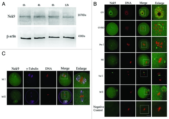
Figure 1. Expression and subcellular localization of Nek9 during mouse oocyte meiotic maturation. (A) Expression of Nek9 was detected by western blotting. Samples were collected after culture for 0, 4, 8 and 12 h, the time points when most oocytes reached the GV, PRO1, MI and MII stages, respectively. The molecular mass of Nek9 and β-actin were about 107 and 43 kDa, respectively. Each sample was collected from 250 oocytes. (B) Subcellular localization of Nek9 as shown by immunofluorescent staining. Oocytes at various stages were stained with an antibody against Nek9 (green); each sample was counterstained with PI to visualize DNA (red). GV, oocytes at germinal vesicle stage; GVBD, oocytes at germinal vesicle breakdown; ProI, oocytes at first prometaphase; MI, oocytes at first metaphase; AnaI, oocytes at first anaphase; TelI, oocytes at first telophase; MII, oocytes at second metaphase. An MII oocyte was used as a negative control for Nek9 confocal microscopy, in which no first antibody was used but the fluorescent second antibody was used. Bar = 20 µm (C) Colocalization of γ-tubulin and Nek9 in GVBD, ProI, MI and MII stages. Oocytes cultured for 8 h (MI) and 12 h (MII) were fixed and stained for γ-tubulin (pink), Nek9 (green) and DNA (red). Bar = 20 µm.

An official website of the United States government
Here's how you know
Official websites use .gov
A
.gov website belongs to an official
government organization in the United States.
Secure .gov websites use HTTPS
A lock (
) or https:// means you've safely
connected to the .gov website. Share sensitive
information only on official, secure websites.
