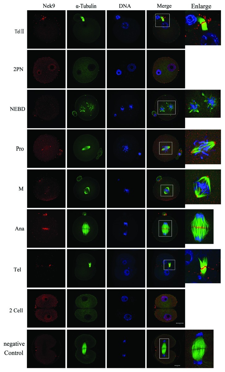
Figure 2. Subcellular localization of Nek9 during fertilization and early embryo cleavage. Subcellular localization of Nek9 during fertilization and early embryo cleavage by immunofluorescent staining. Oocytes at various stages were stained with an antibody against Nek9 (red), α-tubulin (green) and DNA (blue). TelII, zygote at telophaseII stage; NEBD, zygote with one nuclear envelope breakdown (NEBD); Pro, zygote at the stage of pro-metaphase; M, zygote at the stage of metaphase; Ana, zygote at the stage of anaphase; Tel, zygote at the stage of telophase; two-cell embryo. A zygote at the anaphase stage was used as a negative control for confocal microscopy, in which no first antibody was used but the fluorescent second antibody was used. Bar = 20 µm.

An official website of the United States government
Here's how you know
Official websites use .gov
A
.gov website belongs to an official
government organization in the United States.
Secure .gov websites use HTTPS
A lock (
) or https:// means you've safely
connected to the .gov website. Share sensitive
information only on official, secure websites.
