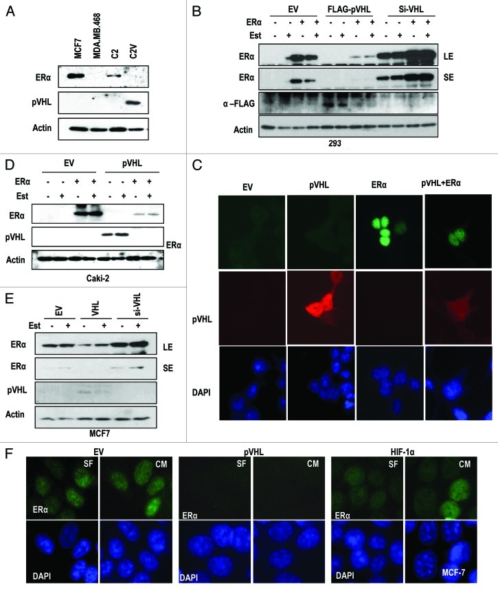
Figure 1. pVHL suppresses ER-α. (A) Expression of ER-α in VHL-deficient C2 cells. MCF7 and MDA-MB.468 (MDA) cells were used as positive and negative controls of ER-α expression. Actin was used as a loading control. (B) pVHL suppresses ER-α. After transfection with the indicated vectors or si-RNAs for 24 h, 293 cells were treated with estrogen (Est; 600 μg/ml) for 6 h. The expression of ER-α was determined by an ER-α-specific Ab, and the expression of exogenous pVHL was determined by a FLAG-Ab. EV indicates an empty vector transfection. LE and SE indicate a long exposure and a short exposure, respectively. (C) The reduction of ER-α and pVHL. Two hundred and ninety-three cells were transfected with VHL and/or ER-α for 24 h. After fixation with Me-OH, 293 cells were stained with anti-ER-α (green), anti-pVHL (red) and DAPI (blue). (D) pVHL suppresses ER-α in pVHL-mutant Caki-2 cells. Caki-2 cells were transfected with the indicated vectors for 24 h and were treated with estrogen for 6 h. (E) pVHL suppresses endogenous ER-α. MCF7 cells were transfected with the indicated vectors or si-RNAs for 24 h and were treated with estrogen for 6 h. LE and SE indicate a long exposure and a short exposure, respectively. (F) The reduction of endogenous ER-α by VHL transfection. MCF-7 cells were transfected with VHL or HIF-1α for 24 h. After fixation with Me-OH, MCF7 cells were stained with anti-ER-α (green) and DAPI (blue). To check for any effects that could have been caused by the serum, cells were incubated in serum-free medium (SF) or complete medium (CM) for 6 h before harvest.

An official website of the United States government
Here's how you know
Official websites use .gov
A
.gov website belongs to an official
government organization in the United States.
Secure .gov websites use HTTPS
A lock (
) or https:// means you've safely
connected to the .gov website. Share sensitive
information only on official, secure websites.
