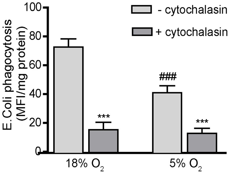
Figure 5. Oxygen tension significantly influences phagocytosis in PMA-differentiated THP-1 cells.

Undifferentiated THP-1 cells were synchronized by serum deprivation for 48 h, plated at a density of 105cells/well in a 96-well plate and differentiated with PMA (20 ng/ml) for 48 h in the absence of 2-ME and FBS. Differentiated THP-1 cells were washed and then incubated for 3 h with E.coli BioParticles®, which emit fluorescence upon acidification in lysosomes following phagocytosis. Phagocytosis, which was quantified by determining the fluorescence intensity at 600 nm, was blocked by pretreating cultures with cytochalasin D (2 µM) for 1 h prior to addition of E. coli BioParticles®. The mean fluorescence intensity was normalized to protein concentration as determined using the BCA protein assay. Data are presented as the mean ± SEM (n = 3 independent experiments). *Significantly different from control (– cytochalasin) treatment under the same oxygen tension; #significantly different from the same culture condition in the 18% O2 treatment group (e.g., 18% O2 versus 5% O2) by Student’s t-test. ***, ### p<0.001.
