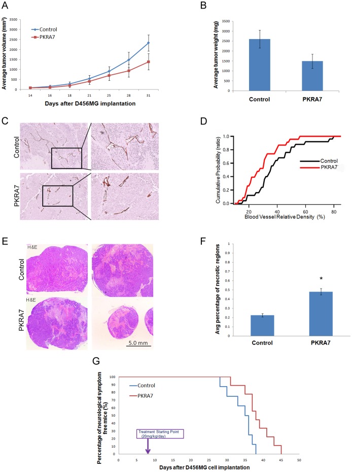
Figure 1. PKRA7 decreases subcutaneous and intracranial glioblastoma xenograft tumor growth.
(A) D456MG cells were SC injected into nude mice, and control (n = 5) or PKRA7 (n = 5) treatment was commenced when tumors became visually detectable (14 days). Measurements were taken every 2–3 days. (B) Average tumor weight of control and PKRA7-treated mouse tumors after removal. (C) IHC staining using CD34 endothelial cell marker in D456MG SC tumors from mice treated with control or PKRA7. (D) Cumulative probability of vessel relative density as measured by CD34 staining. Vascular density of tumors decreased with PKRA7 treatment. (E) Representative pictures of H&E staining of sections from control and PKRA7-treated SC tumors (F) Quantification of necrotic regions from 5 slides of each tumor per treatment group, percentages of necrotic areas were measured by ImageJ (*p≤0.05). (G) 1×10⁴ D456MG cells were IC injected into nude mice and treatment started 7 days after tumor implantation. Mice in control (n = 8) or PKRA7 treatment (n = 9) group were sacrificed when they developed severe neurological phenotype indicative of tumor growth intracranially.

