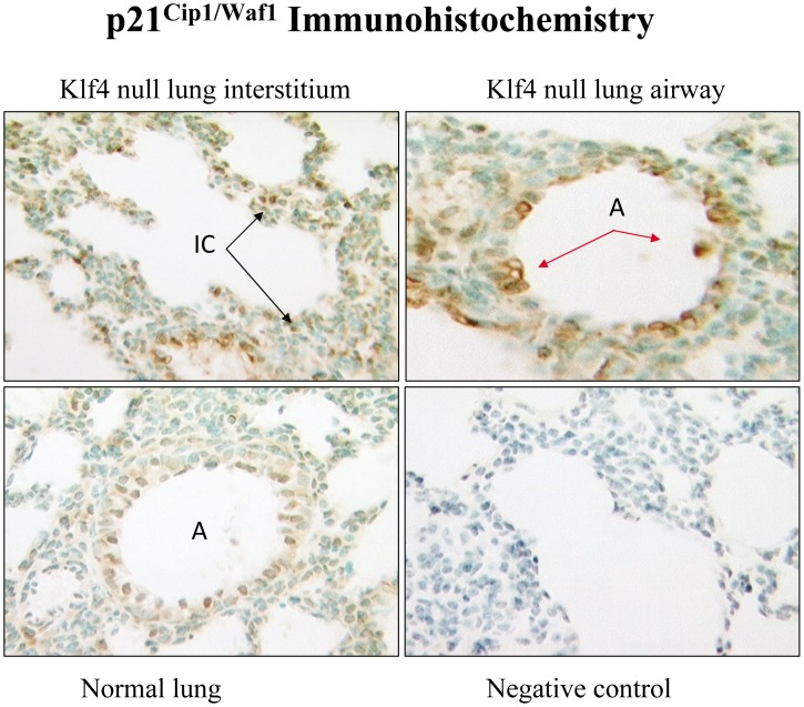
Figure 11. Immunohistochemisry for p21cip1/Waf1 protein.
An intense nuclear immunohistochemical signal (brown color) for p21cip1/Waf1 protein is present in numerous interstitial (IC) cells (top left panel) and airway epithelial (A) cells (top right panel) of Klf4 null lung at birth. Signal is sparse in normal lung (bottom left panel) and absent with omission of primary antibody as the negative control (bottom right panel).

