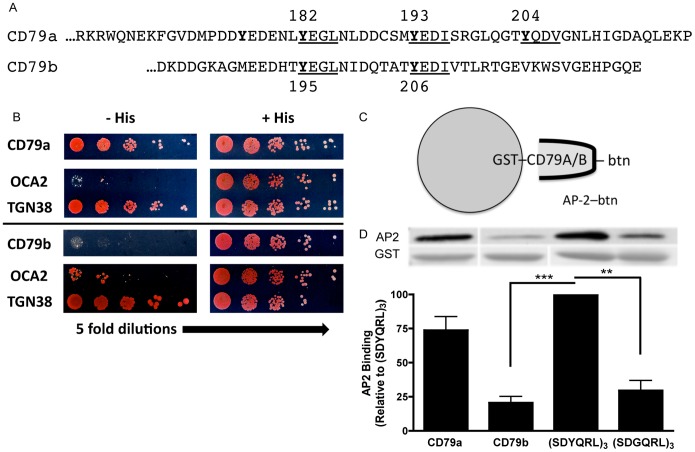
Figure 3. AP2µ Binds to Isolated CD79a but not CD79b.
Panel A, Amino acid sequences of CD79a and CD79b cytoplasmic domains. YxxØ putative AP2 binding motifs underlined. Panel B, AP2µ expressed as a Gal4 activation domain fusion protein was assayed for specific interaction with the cytoplasmic domain of either CD79a or CD79b fused to the Gal4 DNA binding domain. Growth on histidine deficient (His-) plates indicates an AP2–CD79 interaction. The cytoplasmic domain of TGN38 contains a known AP2 binding YxxØ motif and served as a positive control, while the cytoplasmic domain of OCA2 contains a dileucine motif (which does not bind AP2µ) and served as a negative control. Data are representative of 2 experiments. Panel C, Diagram of the GST-CD79 cytoplasmic domain–AP2µ direct binding assay. Panel D, The cytoplasmic domains of CD79a and CD79b were expressed as GST fusion proteins in BL21 E. coli cells. GST-fusion proteins were captured from cell lysates on glutathione beads and the resulting matrix was tested for binding to in vitro translated, biotin-labeled AP2µ. The AP2 binding motif from TGN38, (SDYQRL)3, and a non-AP2-binding derivation containing a tyrosine to glycine substitution, (SDGQRL)3, fused to GST served as positive and negative controls, respectively. Binding is expressed as a percentage of (SDYQRL)3–AP2 interactions. Data is the mean of 3 independent experiments ± S.E.M. Statistical comparisons were measured between SDYQRL and other samples.

