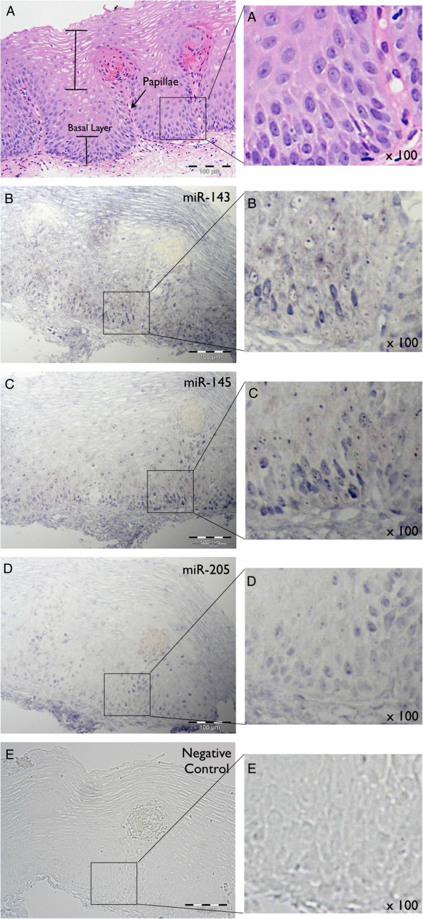
Figure 1.
miRNA in situ hybridisation analysis in oesophageal mucosal biopsies from patients with ulcerative oesophagitis. A: Staining with Haematoxylin and Eosin. The basal layer, papillae and differentiated squamous epithelium are clearly visible. B, C, D and E: Hybridisation with LNA-probes for miRNAs miR-143 (B), miR-145 (C) and miR-205 (D), and LNA-negative control (E). No hybridisation was observed with the LNA-negative control probe. A region from each section has been magnified 100x. In A, rounded nuclei in the oesophageal epithelium are evident. B, C &D depict the punctate nuclear staining observed for each miRNA.

