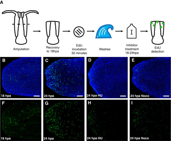
Figure 4.
Efficacy of hydroxyurea and nocodazole in blocking cell proliferation. (A) Schematic of experiments. (B-E) Nuclei of proliferating cells (green) labeled with the thymidine analog EdU, and all nuclei counterstained with Hoechst (blue). (F-I) Nuclei of proliferating cells labeled with EdU (green). (B, F) Region of the wound site in a polyp incubated with EdU for 30 minutes at 18 hpa, and fixed immediately after EdU incubation for detection. (C, G) Region of the wound site in a polyp incubated with EdU for 30 minutes at 18 hpa, washed and maintained in 1/3x filtered seawater until 24 hpa, and fixed 24 hpa. (D, H) Region of the wound site in a polyp incubated with EdU for 30 minutes at 18 hpa, washed and maintained in 1/3x filtered seawater with 20 mM hydroxyurea until 24 hpa, and fixed 24 hpa. (E, I) Region of the wound site in a polyp incubated with EdU for 30 minutes at 18 hpa, washed and maintained in 1/3x filtered seawater with 0.1 μM nocodazole until 24 hpa, and fixed 24 hpa. Scale bars = 50 μm.

