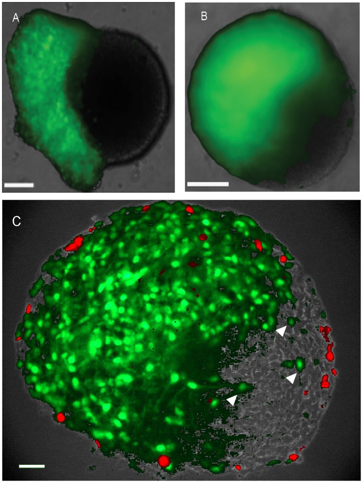
Figure 2. Cell migration and proliferation in chimeric mouse neurospheres.
eGFP expressing neurosphere cells were centrifuged onto unlabeled intact neurospheres to produce chimeric neurospheres. Wholemount fluorescence images of living neurospheres taken (A) after 24 h and (B) after 96 h culture, showing green eGFP fluorescence: note that after 96 h the boundary between labeled and unlabeled cells has become diffuse. C, 8 µm equatorial section of typical chimeric neurosphere after 96 h culture, at the end of which the neurospheres had been incubated with 10 µM BrdU for 1 hour before fixation. Note that eGFP cells have migrated into the unlabeled half of the chimeric neurosphere (C, arrow heads), and that BrdU labelling (red) is restricted to the periphery of the chimeric neurosphere. Scale bars = 100 µm.

