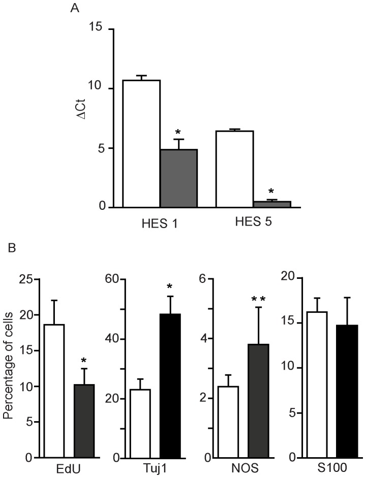
Figure 5. Effects of DAPT-mediated γ-secretase inhibition on cell proliferation and expression of neuronal and glial markers by mouse neurosphere cells.
After 2 weeks culture mouse neurospheres were cultured for a further 96 h in the presence of 20 µM DAPT (shaded columns) or DMSO solvent control (clear columns). A: Levels of Hes1 and Hes5 mRNA determined by q-PCR. Columns show the ΔCt values, normalised to β-actin levels (± SEM, means of 3 individual experiments). B: Expression of neuronal and glial markers. Cells dissociated from the neurospheres were allowed to adhere to Permanox slides before fixation and staining for EdU, Tuj1, NOS and S100. Nuclei were counter-stained with DAPI. Fluorescent cells were counted in 5 random optical fields in each chamber using a 40 x oil objective. Error bars are ± SEM, (values from 3–5 experiments). A two-tailed t-test was performed for differences between open and closed columns for each marker. *p<0.01.

