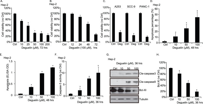
Figure 1. Cytotoxic effects of deguelin in cultured HNSCC cells.
Hep-2 cells were exposed to deguelin at a various concentration (10, 25, 50, 100 and 200 µM) for 72 hours (A) or exposed to 100 µM of deguelin for different time points (12, 24, 48 and 72 hours) (B), cell viability was then measured by MTT assay. (C) Three different cell lines A253, SCC-9 and PANC-1 were exposed to 100 µM of deguelin (Deg) for 72 hours; cell viability was measured by MTT assay. Hep-2 cells were treated with indicated concentration of deguelin, hoechst nuclear staining and enzyme-linked immunosorbent cell apoptosis assay were utilized to analyze cell apoptosis (D–E), caspase-3 activity was also measured (F), apoptosis related proteins including cleaved caspase-3, cleaved caspase-9, Bcl-Xl were detected by Western blots (G), Bcl-Xl expression level was quantified using image J software (H). The mean of at least three independent experiments performed in triplicate is shown. Statistical significance was analyzed by ANOVA. *p<0.01 vs Control.

