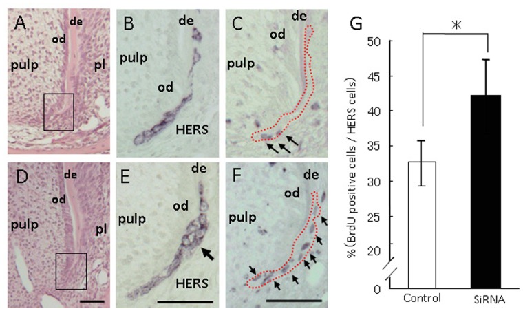
Figure 4. Light micrographs illustrating HERS treated with siRNAs.
AMBN siRNA injected mice revealed a multilayered appearance in the basal portion of HERS and increased BrdU positive cells in that of the outer layer. Treatment with control siRNA (A–C) and AMBN siRNA (D–F). (A) and (D) Hematoxylin-eosin staining. (B) and (E) Immuno-staining with cytokeratin 5. Treatment with AMBN siRNA revealed a multilayered appearance in the basal portion of HERS (arrow). (C) and (F) Immuno-staining with AMBN. Treatment with AMBN siRNA increased BrdU positive cells in the outer layer of HERS (arrows). de, dentin; od, odontoblasts; pl, periodontal ligament; HERS, Hertwig’s root sheath. Red dotted lines mark the location of HERS. Scale bars: 100 µm. (G) The percentage of BrdU positive cells per total HERS cells was counted and compared between treatments with control and AMBN siRNA. The percentage of BrdU positive cells was higher with treatment with AMBN siRNA (n = 10) than that with control siRNA (n = 10). Statistical analysis was performed using Student’s t-test, *; p<0.05.

