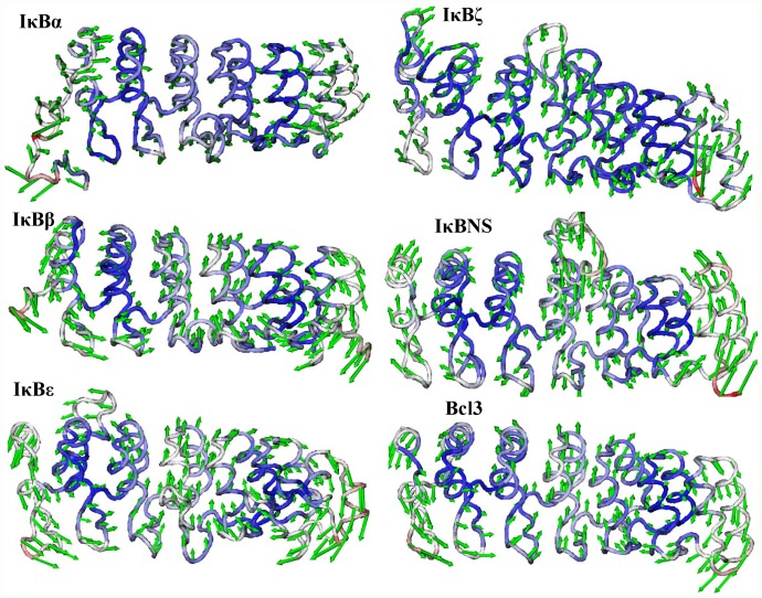
Figure 9. Lowest frequency mode 3 obtained for IκB proteins using ANM.
The lowest frequency mode 3 results are shown from top to bottom. Only the Cα atoms are shown for clarity. The atoms in red indicate large fluctuations, whereas the blue color corresponds to small fluctuations. The magnitude and direction of the displacement for mode 3 are represented by the green arrows.

