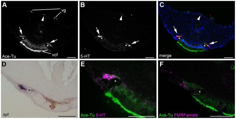
Figure 3. Ventral and dorsal nerve cords in the vestimental region.
A–C, E: Anti-acetylated tubulin and anti-serotonin immunoreactivities. Serotonergic neural cell bodies are located on the lateral side of the giant axon (asterisks), and they project their axons medially (arrows). Anti-serotonin immunoreactivity was found in the dorsal nerve cords (arrowheads). D: Expression of syt was detected in the ventral nerve cord. F: Double-stain against anti-acetylated tubulin (green) and anti-FMRFamide (magenta) antibodies shows the presence of FMRFamidergic neurons in the ventral nerve cord. Scale bars = 100 µm. vcf, ventral ciliated field; vg, vestimental groove.

