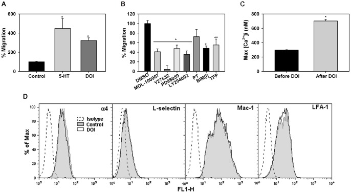
Figure 6. 5HT and DOI induce migration of murine Eos.
(A) Migration of murine Eos towards 10 μM 5-HT or DOI in Transwell® plates after 4 h at 37°C. The average number of cells/field/well was determined and results expressed as a percentage of background migration observed in wells containing medium alone. Combined data (Mean ± SEM) of 5 independent experiments is shown. *p<0.01 for comparison of 5-HT- or DOI-treated cells versus vehicle-treated cells. (B) Effect of MDL-100907, Y27632, PD98059, LY294002, BIM(I), TFP (all at 10 μM), and PT (100 ng/ml) on DOI-induced migration of murine Eos. Cells were pre-treated with inhibitors or DMSO (vehicle) alone for 20 min before addition to Transwell® Chambers. Results are expressed as a percentage of the migration of vehicle treated cells towards DOI. Combined data (Mean ± SEM) of 3 (for PT, BIM(I), TFP) or 5 (all other inhibitors) independent experiments in duplicate or triplicate is shown. *p<0.01 and **p<0.02 for comparison of cells treated with inhibitors versus cells treated with vehicle. (C) Basal and DOI-induced [Ca2+]i levels in murine Eos from 364 cells by digital videofluorescence imaging with Fura-2 AM. Representative data of three independent experiments performed in triplicate. *p<0.01 compared to unstimulated cells. (D) Expression of adhesion molecules by murine Eos after treatment with 10 μM DOI (or PBS) for 5 min by flow cytometry using rat mAbs against α4 (CD49), Mac-1 (CD11b) and LFA-1 (CD11a) followed by FITC-conjugated goat anti-rat IgG as the secondary antibody. Depending on the mAb, rat IgG2a or 2b was used as the isotype matched control. Expression of CD62L was evaluated using FITC-conjugated anti-mouse CD62L (BD Biosciences). FITC-conjugated rat-IgG2a was used as the isotype control for CD62L. All antibodies were used at a final concentration of 5 μg/ml. Data shown is representative of three independent experiments with Eos from different mice.

