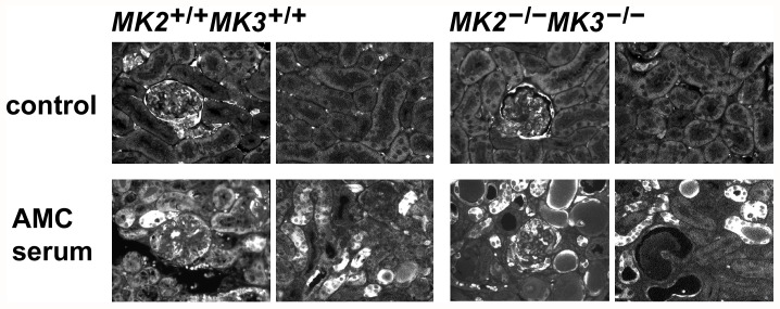
Figure 6. Distribution of HSPB1 in renal cortices in response to the AMC serum.
Paraffin-embedded renal cortices of untreated and AMC serum-treated wild-type and MK2/MK3 double knock-out mice (day 16 following AMC serum treatment) were sectioned and processed for immunofluorescence microscopy. Total HSPB1 was visualized using an anti-HSPB1 antibody. In untreated control mice of either genotype, labeling of the glomeruli (including Bowman's space) was moderately elevated as compared to the surrounding tubules (upper row, left panels) or to the more distant tubules (upper row, right panels). AMC serum treatment caused a strong increase in HSPB1 labeling in the tubules, both adjacent to the glomeruli (lower row, left panels) and more distant from the glomeruli (lower row, right panels), thus indicating a stress response in the tubular compartment.

