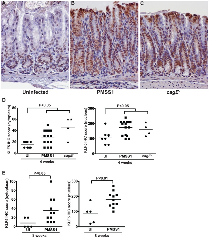
Figure 4. H. pylori upregulates KLF5 expression in vivo.
(A–C) KLF5 expression in murine antral gastric tissue was assessed by KLF5 immunostaining in uninfected (A), H. pylori PMSS1-infected mice (B), and H. pylori PMSS1 cagE−-infected mice (C) at 400× magnification. (D and E) A single pathologist, blinded to treatment groups, assessed and scored KLF5 immunostaining. KLF5 immunohistochemistry (IHC) score was determined by assessing the percentage of KLF5+ epithelial cells multiplied by the intensity of epithelial KLF5 staining (1–3) in both the cytoplasm and nucleus of murine gastric epithelial cells (D and E). Each data point represents an individual animal and mean values are shown. Circles designate uninfected mice, squares represent H. pylori PMSS1-infected mice, and triangles represent H. pylori PMSS1 cagE−-infected mice. Mann-Whitney and ANOVA tests were used to determine statistical significance between groups.

