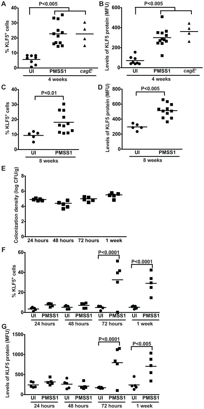
Figure 5. H. pylori induces expansion of a KLF5+ cell population in vivo.
(A–G) KLF5 expression in murine gastric epithelial cells was assessed by flow cytometry analysis in uninfected and H. pylori-infected mice at acute time points (24, 48, 72 hours, and 1 week) and chronic time points (4 and 8 weeks) post-challenge. Percentage of KLF5+ cells at 4 weeks (A) and 8 weeks (C) and levels of KLF5 protein at 4 weeks (B) and 8 weeks (D), as determined by mean fluorescence units (MFU), were determined by flow cytometry. Data from 4 and 8 week time points were analyzed at separate times. H. pylori colonization density in mice infected for 24, 48, and 72 hours, and 1 week was assessed by quantitative culture (E). Percentage of KLF5+ cells (F) and levels of KLF5 protein (G) at 24, 48, or 72 hours, or 1 week were determined by flow cytometry. Each data point represents gastric epithelial cells analyzed from a single animal and mean values are shown. Circles designate uninfected mice, and squares represent H. pylori-infected mice. Mann-Whitney and ANOVA tests were used to determine statistical significance between groups.

