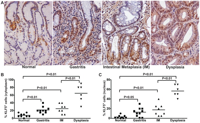
Figure 8. KLF5 expression parallels the severity of gastric premalignant lesions in H. pylori-infected humans.
(A) KLF5 expression was evaluated by immunohistochemistry in a human population at high risk for gastric cancer. Gastric biopsies from uninfected patients with normal gastric mucosa and H. pylori-infected patients with non-atrophic gastritis, intestinal metaplasia (IM), and dysplasia were evaluated for KLF5 immunostaining at 200× magnification. (B and C) A single pathologist assessed the percentage of KLF5+ cells exhibiting cytoplasmic (B) or nuclear (C) staining. Each data point represents an individual biopsy and mean values are shown. The percentage and mean value of KLF5+ cells from biopsies from patients with normal gastric tissue (circles), gastritis (squares), intestinal metaplasia (IM, triangles), and dysplasia (inverted triangles) are shown. Mann-Whitney and ANOVA tests were used to determine statistical significance between groups.

