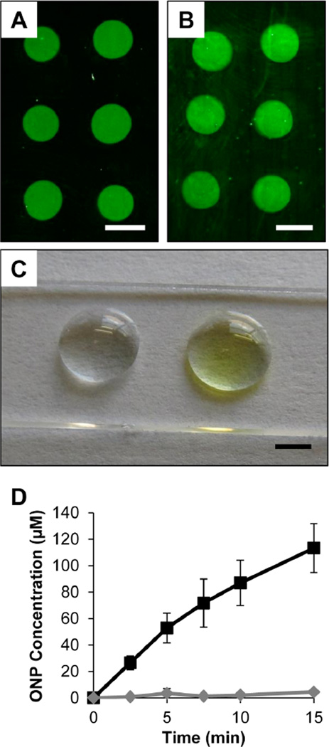
Figure 4.
(A–B) Representative fluorescence micrographs of glass substrates coated with PEI/PVDMA films spotted with (A) FITC-labeled streptavidin or (B) unlabeled streptavidin (the image in (B) was treated with anti-HA-biotin and anti-rat IgG Alexa Fluor 488 prior to imaging; see text). (C) Digital photograph of film-coated glass containing spots of immobilized β-galactosidase (right spot) or BSA (left spot) incubated under droplets of ONPG for 10 minutes. (D) Plot of ONP concentration vs. time measured from a droplet incubated on a spot containing immobilized β-galactosidase (black squares) or BSA (grey diamonds). Scale bars are (A–B) 1 mm and (C) 2 mm.

