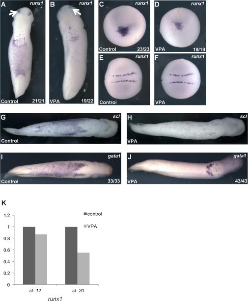
Figure 2. VPA inhibits the development of erythroid progenitors.
A) Primitive erythroid marker runx1 is expressed within the VBI and olfactory placode (arrows) in control early tailbud (stage 26) embryos. B) VPA reduced runx1 expression in the VBI. C) In control late neurulae (stage 19, anterior view, with dorsal oriented to the top), runx1 is expressed in precursors of the aVBI, which lie in a ventral-anterior domain at this stage. D) VPA markedly reduces runx1 expression within presumptive hematopoietic cells in neurula stage embryos (same orientation as panel C). E) and F) Dorsal view (anterior to the left)- Expression of runx1 in Rohan Beard cells in the same embryos as (C) and (D) is not affected by VPA. G-J) Ventral views, anterior to the right. VPA treatment (H) and (J) reduces expression of erythroid markers scl (G) and (H) and gata1 (I) and (J) in the VBI of tailbud embryos (stage 31). K) runx1 expression in whole embryos is reduced with VPA treatment at late neurula stage (stage 18), as determined by qRT-PCR (data represent the mean of 4 independent experiments, normalized to ODC).

