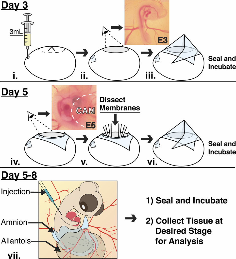
Fig. 1.
A schematic diagram showing treatment of the eggs and embryos at different stages of development. (i–iii) After 3 days of incubation, albumen is withdrawn from the narrow end of the egg, which is then windowed to reveal an intact E3 chick embryo and surrounding vasculature. The eggs are sealed with transparent tape and re-incubated. (iv–vi) On day 5, the tape is cut to reveal the E5 embryo that is partially covered by the CAM. The amnion is dissected and the CAM is displaced away to expose the embryo. The exposed embryo can be manipulated at this stage or re-incubated if later stages are desired. (vii) The exposed embryos do not show any defects and are accessible in ovo between E5–E8 and they can be manipulated and re-incubated until a desired time point.

