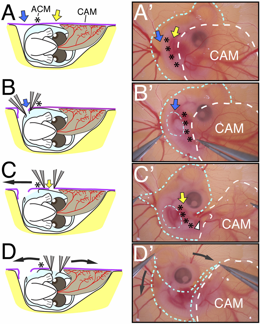
Fig. 2.
Exposure of the E5 chick embryo in ovo. (A–D) Schematics of the embryo viewed from the posterior showing the surrounding amnion (light blue), the chorion (purple) and the vascularized allantois (brown) during membrane dissections. (A’–D’) Bright-field images of the embryo showing the location of the amnion (blue outline) and chorioallantoic membrane (white outline) during membrane dissections. The amnion and chorion are dissected as indicated by the blue and yellow arrows in the regions lateral to the ACM (asterisks). Following the separation of the embryo and surrounding amnion from the CAM (C’), the amnion is peeled away from the cranial region (D’, curved black arrows). The posterior region of the embryo slides out of the amnion and the entire embryo becomes exposed.

