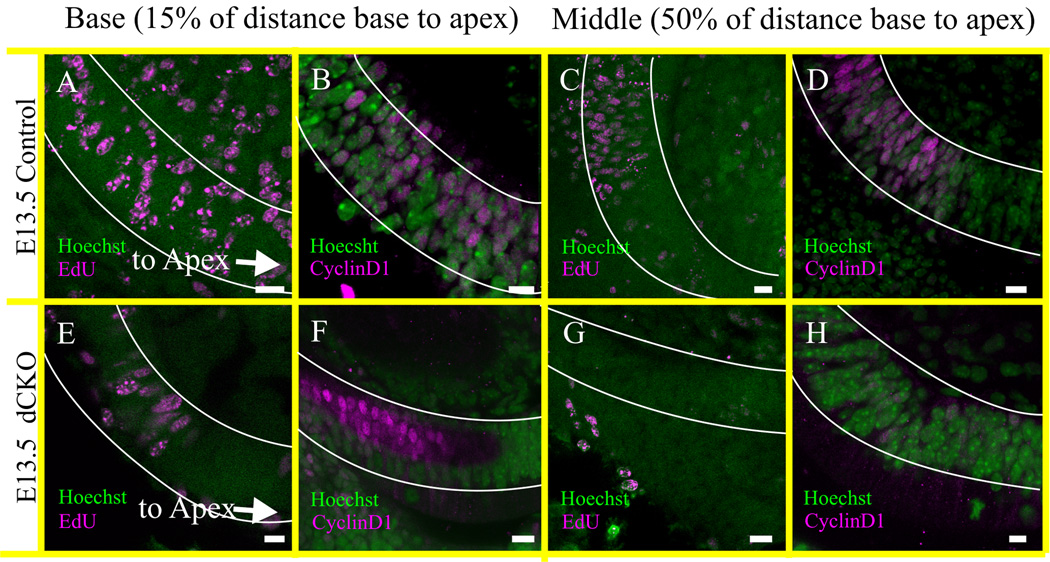
Figure 6. Proliferation ends prematurely in the cochlea of dCKO mice.
The proliferation of the organ of Corti is assessed at E13.5 by the injection of EdU eight hours before collection at E13.5 (A,E,C,G). EdU positive staining is indicative of DNA synthesis and the proliferating cells are in the S phase of the cell cycle. The proliferation detected with the EdU is confirmed by the CyclinD1 immunohistochemistry at E13.5 mice (B,F,D,H) and both are co-labeled with the Hoechst nuclear stain (A–H). The proliferating cells are reduced in the ‘basal part’ of cochlea in the dCKO compared to control littermate shown by both EdU and CyclinD1 staining (compare A,B with E,F). However, in the ‘middle’ part of the cochlea, no EdU or CyclinD1 positive cells are detected in the dCKO mice (compare C,D with G,H). EdU and CyclinD1 positive cells are reduced in the ‘middle’ part of the control cochlea than that of the ‘base’ indicating a natural decline of proliferation by E13.5 which usually starts from apex at E12 (A–D) (Matei et al., 2005). In contrast, complete absence of EdU and CyclinD1 positive cells at ‘middle’ and reduction at ‘base’ already by E13.5 suggests the premature cessation of proliferation in the dCKO cochlea (E–H). The distribution of these cells are scored as ‘base’ if it was at 15% of the distance from the basal tip of the organ of Corti (outlined in white; A,B,E,F), and as ‘middle’ if it was near 50% of the distance between base and apex (outlined in white; C,D,G,H). Proliferation is not assessed in the vestibular organs. Scale bar = 10 µm.

