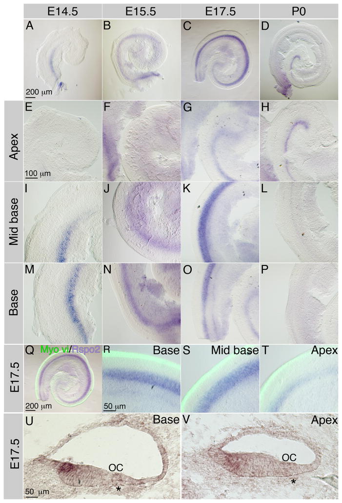
Figure 1.
Spatial and temporal expression of Rspo2 in the developing cochlea. Low magnification views of whole mount in situ hybridization for Rspo2 at E14.5 (A), E15.5 (B), E17.5 (C) and P0 (D) show that Rspo2 is first expressed in the base at E14.5 (A), extended to the mid base by E15.5 (B), and the apex by E17.5 (C). At P0, expression of Rspo2 is maintained in the apex but was down regulated in the base. E–H high magnification views of the least developmentally advanced region of the cochlea, the apex, at E14.5 (E), E15.5 (F), E17.5 (G) and P0 (H) show that Rspo2 was not detected in the apex until E17.5, relatively late in cochlear development. I–L show high magnification views of the mid base of the cochlea at E14.5 (I), E15.5 (J), E17.5 (K) and P0 (L) show that the Rspo2 expression domain rapidly extends along the mid base from E14.5 (I), peaking at E17.5 (K), but is beginning to be down regulated by P0. M–P show high magnification views of the most mature region of the cochlea, the base, at E14.5 (M), E15.5 (N), E17.5 (O) and P0 (P). Rspo2 expression was detected strongly in the base at E14.5 (M) and E15.5 (N) but by E17.5, relative to its expression in the mid base, Rspo2 expression was starting to be down regulated. By P0, there was no Rspo2 expression in the base. Q The whole mount E17.5 cochlea shown in panels C, G, K and O showing in situ hybridization for Rspo2 (purple) and immunofluorescence for Myosin vi (green). R–T show higher magnifications of the base ©, mid base (S) and Apex (T) of the cochlea shown in Q/C. In the base and mid base, The Rspo2 domain was situated relatively far from the organ of Corti (labeled with Myosin vi) (Q, R, S). In the apex, Myosin vi labeling showed that the inner hair cells began to differentiate before Rspo2 expression reached levels detectable by in situ hybridization. U. In situ hybridization for Rspo2 on a transverse section through the basal region of an E17.5 cochlea shows Rspo2 expression in the luminal surface cells of the most lateral region of the GER. V. In situ hybridization for Rspo2 on a transverse section through the apical region of an E17.5 cochlea shows that in a less mature region of the cochlea, expression spans the depth of the GER and is more diffuse. * indicate the spiral vessel – a marker for the location of the developing organ of Corti (OC).

