Figure 5.
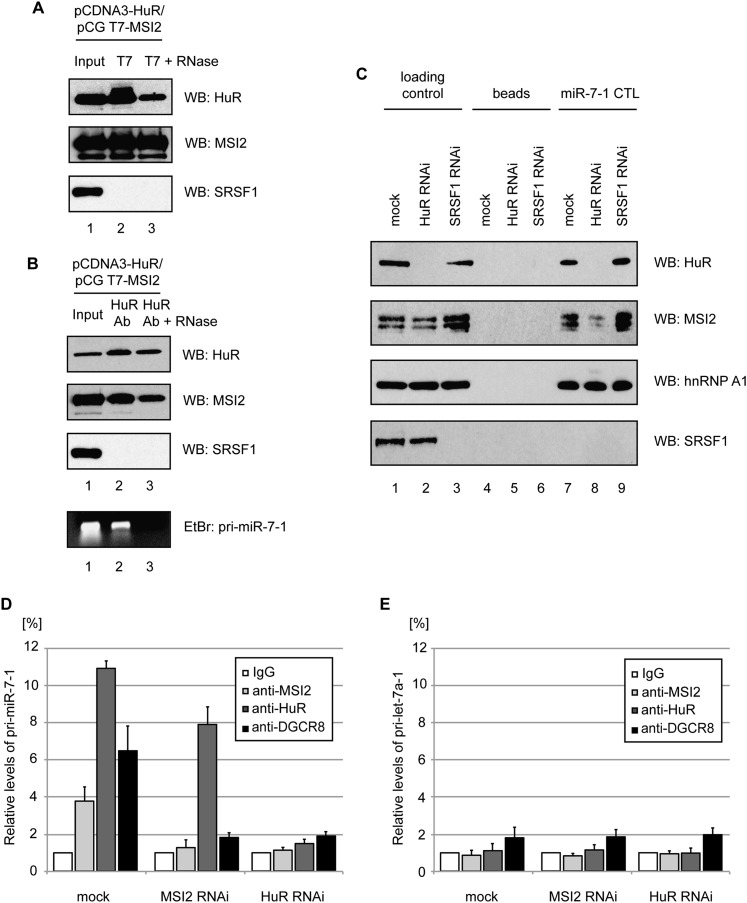
HuR recruits MSI2 to the pri-miR-7-1 CTL through a direct interaction. (A) Extracts prepared from HeLa cells transfected with pCG T7-MSI2 and pCDNA3-HuR were incubated with T7 agarose. (Lane 2) The bound proteins were separated on a 10% SDS–polyacrylamide gel and analyzed by Western blotting with anti-HuR, anti-MSI2, or anti-SRSF1 antibodies. (Lane 3) Alternatively, the immunoprecipitate was treated with RNases A/T1 prior to loading on the gel. Lane 1 was loaded with 2% of the amount of extract used for each immunoprecipitation. (B) Extracts prepared from HeLa cells transfected with pCG T7-MSI2 and pCDNA3-HuR were incubated with protein A beads and anti-HuR antibody. (Lane 2) The bound proteins were separated on a 10% SDS–polyacrylamide gel and analyzed by Western blotting with anti-HuR, anti-MSI2, or anti-SRSF1 antibodies. (Lane 3) Alternatively, the immunoprecipitate was treated with RNases A/T1 prior to loading on the gel. Lane 1 was loaded with 2% of the amount of extract used for each immunoprecipitation. Ethidium bromide-stained 1% agarose gel of RT–PCR assay detecting pri-miR-7-1 in input; anti-HuR immunoprecipitation and anti-HuR immunoprecipitation with RNase treatment are shown in lanes 1–3, respectively. (C) Western blot analysis of miR-7-1 CTL RNA pull-down in mock-, HuR- or SRSF1-depleted HeLa cell extracts for HuR, MSI2, hnRNP A1, and SRSF1. Lanes 1–3 show loading control of HeLa cell extracts from mock, HuR RNAi, and SRSF1 RNAi, respectively. Lanes 4–6 show reactions with beads only. Lanes 7–9 represent miR-7-1 CTL RNA pull-down in HeLa cell extracts from mock, HuR RNAi, and SRSF1 RNAi, respectively. (D,E) RNA immunoprecipitation assays of formaldehyde cross-linked HeLa cells using anti-MSI2, anti-HuR, and anti-DGCR8 antibodies, coupled with protein-A agarose beads. qRT–PCR analysis was performed on TRIzol LS-isolated RNA with primers detecting pri-miR-7-1 (D) and pri-let-7a-1 (E) transcripts. The percentage of immunoprecipitation was plotted relative to values derived from IgG controls, which were set to 1. Mean values and SDs of three independent experiments are shown.

