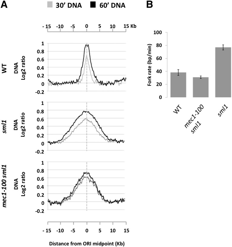
Figure 6.
Replication fork rate is compromised in mec1-100 cells in HU. (A) Average DNA profiles at 30 min (gray line) and 60 min (black line) from early efficient ORIs (n = 8) in wild type (WT), sml1, and mec1-100 sml1 cells. Data were smoothed using a moving average window of 800 bp. (B) Average replication fork rates from two independent biological replicates measured from 30 to 60 min in 200 mM HU in the same strains as in A. Error bars indicate the standard error of the mean.

